It's not a question I get asked everyday. But sheep do have a reputation for dying for no reason whatsoever. That's not a view I subscribe to, there's obviously a reason behind every loss, so learning more is important for the health of our flock.
Post Mortem Evidence Collection as an Aid to Veterinary Diagnosis of Sheep Diseases. That was the snappy title given to the six hour course run by a veterinary surgeon. We began with a powerpoint presentation that set out the basics of sheep anatomy and why an autopsy performed on farm is worthwhile.
Farmers look at their stock everyday, and you can notice characteristics and behaviours that point toward ill health. We were advised to consider the following list:
1. Weight loss
2. Respiratory signs
3. Scour (farm/vet language for diarrhoea)
4. Abortion
5. Neurological
6. Skin / Wool
7. Lameness
8. Sudden death!
Clearly the latter sign would tell you something is definitely wrong! But what..? In the lead up to death had the animal displayed any of the other signs? Observation at every stage is important, but eventually the only way to really tell what happened is to look inside. In all cases the autopsy had to be conducted within 24 hours of death.
Allowing farmers to conduct their own investigations isn't as precise as the service offered by the Veterinary Laboratories Agency (the VLA - our nearest is in Thirsk), but it does offer an instant answer for diseases with specific pathology e.g. fluke (see: http://en.wikipedia.org/wiki/Trematoda). That rapid determination then allows for wider flock treatment decisions.
As well as speed another consideration is cost, a VLA examination is £160 including carcass disposal. Clearly a matter requiring some thought balanced against the risk of potentially not discovering the cause of death. In my case I could see myself examining one or two sheep to find the cause of death but if I failed I would be looking toward our vet and the VLA for professional help, especially if losses were mounting.
We were given a full round up of the kit required, including the sense behind vets wearing a pair of long gloves and another short pair over the top for better feel. The last item on the list was a digital camera.
 These pictures are not taken to shock, but rather as an important personal aide memoire. In addition as clearly as I could describe the colour of specific internal organs that information can provide clues to aid diagnosis.
 First things first, a quick check of the teeth. This ewe was six years old, so it would be normal for her to be missing teeth. The teeth you can't easily see (the mouth can't be opened wide enough) are the molars. In bad condition the sheep would not be able to cud. In this case she appeared to be ok for her age.
The next task was to cut the skin and fleece away with a sharp knife. In doing so we were reminded to only use sharp tools and not to get carried away, our instructor could relate stories involving friends that had suffered injury as a result of being over eager with a blade.
This picture shows the wind pipe. As well as not cutting ourselves we were also told to be careful to avoid damage to the organs and the potential for contamination of any samples.


One of two healthy looking kidneys, see below.


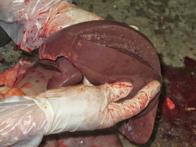
It is amazing to note however that the sheep can survive with up to 70% liver damage and recover after treatment.

In a healthy animal the lungs would be a fluffy pink colour. Here they are clearly not and are burdened with white'ish tumors. Clearly we had identified the problem; the vet diagnosed Maedi Visna (MV).
 Sheep farmers will be familiar with the disease. If you are a regular visitor to agricultural shows over the summer months you may have noticed that sheep are often split into groups, one MV accredited and the other not. MV travels through the air and the sheep are separated to avoid the risk of the disease being transmitted.
A number of this flock are now having their blood tested to check for MV. It may be that this sheep was the only victim. Certainly without the autopsy the illness would not have been identified and the rest of the flock left at risk.
No comments:
Post a Comment